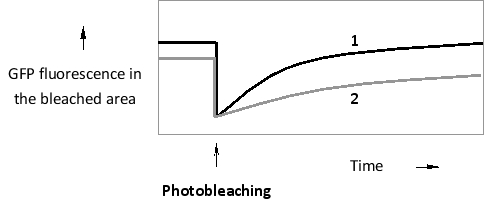
A certain GTP-binding protein can exist in two main states. When bound to GDP, it is mostly cytosolic. In its GTP-bound form, however, it associates with the cytosolic face of the endoplasmic reticulum (ER) membrane, where it hydrolyzes the bound GTP after a short delay and is released again into the cytosol. You have created and expressed green fluorescent protein (GFP) fusions of the wild-type protein, as well as that of a mutant protein that does not bind GTP as readily as the wild type. You then perform a fluorescence recovery after photobleaching (FRAP) experiment by photobleaching a small area of the ER membrane and measuring GFP fluorescence recovery over time. According to the results below, which curve (1 or 2) do you think corresponds to the wild-type fusion protein? Write down 1 or 2 as your answer.
Correct Answer:
Verified
View Answer
Unlock this answer now
Get Access to more Verified Answers free of charge
Q10: Two segments (S1 and S2) in a
Q11: What is the advantage of using quantum
Q12: Given the absorption and emission spectra of
Q13: Which microscopy set-up uses a longer wavelength
Q14: Atomic force microscopy (AFM) is used in
Q16: Indicate true (T) and false (F) statements
Q17: The light used to excite a fluorescent
Q18: Indicate whether you would use a fluorescent
Q19: Imagine a transcription regulatory protein (X) that
Q20: In the diagram below, a logarithmic scale
Unlock this Answer For Free Now!
View this answer and more for free by performing one of the following actions

Scan the QR code to install the App and get 2 free unlocks

Unlock quizzes for free by uploading documents