What projection and anatomy is demonstrated in the image below? 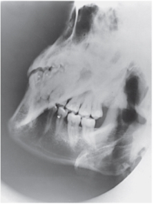
A) AP axial of the TMJs
B) PA axial of the mandibular rami
C) PA of the mandibular body
D) Axiolateral oblique of the mandibular body
Correct Answer:
Verified
Q135: The sinus identified in the figure below
Q136: What projection (method)of the facial bones is
Q137: Which part of the patient's face is
Q138: At which level will the central ray
Q139: What projection and anatomy is demonstrated in
Q141: Which reference line is positioned horizontal to
Q142: At which level should the central ray
Q143: The central ray forms an angle of
Q144: What projection (method)is demonstrated in the image
Q145: The ethmoidal sinuses are located within which
Unlock this Answer For Free Now!
View this answer and more for free by performing one of the following actions

Scan the QR code to install the App and get 2 free unlocks

Unlock quizzes for free by uploading documents