




 Chat with AI


Inquiry into Life 15th Edition by Sylvia Mader ,Michael Windelspecht
Edition 15ISBN: 978-1259426162Inquiry into Life 15th Edition by Sylvia Mader ,Michael Windelspecht
Edition 15ISBN: 978-1259426162 Exercise 2
Modern Microscopy
Microscopes have given scientists a deeper look into how life works than is possible with the naked eye. Today, there are many types of microscopes. A compound light microscope uses a set of glass lenses to focus light rays passing through a specimen to produce an image that is viewed by the human eye. A transmission electron microscope (TEM) uses a set of electromagnetic lenses to focus electrons passing through a specimen to produce an image, which is projected onto a fluorescent screen or photographic film. A scanning electron microscope (SEM) uses a narrow beam of electrons to scan over the surface of a specimen that is coated with a thin metal layer. Secondary electrons given off by the metal are detected and used to produce a three-dimensional image on a television screen. Figure 3A shows these three types of microscopes and their images.
Magnification, Resolution, and Contrast
Magnification is the ratio between the size of an image and its actual size. Electron microscopes magnify to a greater extent than do compound light microscopes. A light microscope can magnify objects about a thousand times, but an electron microscope can magnify them hundreds of thousands of times. The difference lies in the means of illumination. The path of light rays and electrons moving through space is wavelike, but the wavelength of electrons is much shorter than the wavelength of light. This difference in wavelength accounts for the electron microscope's greater magnifying capability and its greater ability to distinguish between two points (resolving power).
Resolution is the minimum distance between two objects that allows them to be seen as two separate objects. A microscope with poor resolution might enable a student to see only one cellular granule, while the microscope with the better resolution would show two granules next to each other. The greater the resolving power, the greater the detail seen.
If oil is placed between the sample and the objective lens of the compound light microscope, the resolving power is increased, and if ultraviolet light is used instead of visible light, it is also increased. But typically, a light microscope can resolve down to 0.2 ?m, while the transmission electron microscope can resolve down to 0.0002 ?m. If the resolving power of the average human eye is set at 1.0, then the typical compound light microscope is about 500, and the transmission electron microscope is 500,000.
The ability to make out, or resolve, a particular object can depend on contrast, a difference in the shading of an object compared to its background. Higher contrast is often achieved by staining cells with colored dyes (light microscopy) or with electron-dense metals (electron microscopy), which make them easier to see. Optical methods such as phase contrast and the use of fluorescently tagged antibodies can also help us visualize subcellular components such as specific proteins.
Figure 3A Diagram of microscopes with accompanying micrographs. The compound light microscope and the transmission electron microscope provide an internal view of an organism. The scanning electron microscope provides an external view of an organism.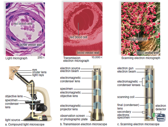
Why is it necessary to artificially color electron micrographs?
Microscopes have given scientists a deeper look into how life works than is possible with the naked eye. Today, there are many types of microscopes. A compound light microscope uses a set of glass lenses to focus light rays passing through a specimen to produce an image that is viewed by the human eye. A transmission electron microscope (TEM) uses a set of electromagnetic lenses to focus electrons passing through a specimen to produce an image, which is projected onto a fluorescent screen or photographic film. A scanning electron microscope (SEM) uses a narrow beam of electrons to scan over the surface of a specimen that is coated with a thin metal layer. Secondary electrons given off by the metal are detected and used to produce a three-dimensional image on a television screen. Figure 3A shows these three types of microscopes and their images.
Magnification, Resolution, and Contrast
Magnification is the ratio between the size of an image and its actual size. Electron microscopes magnify to a greater extent than do compound light microscopes. A light microscope can magnify objects about a thousand times, but an electron microscope can magnify them hundreds of thousands of times. The difference lies in the means of illumination. The path of light rays and electrons moving through space is wavelike, but the wavelength of electrons is much shorter than the wavelength of light. This difference in wavelength accounts for the electron microscope's greater magnifying capability and its greater ability to distinguish between two points (resolving power).
Resolution is the minimum distance between two objects that allows them to be seen as two separate objects. A microscope with poor resolution might enable a student to see only one cellular granule, while the microscope with the better resolution would show two granules next to each other. The greater the resolving power, the greater the detail seen.
If oil is placed between the sample and the objective lens of the compound light microscope, the resolving power is increased, and if ultraviolet light is used instead of visible light, it is also increased. But typically, a light microscope can resolve down to 0.2 ?m, while the transmission electron microscope can resolve down to 0.0002 ?m. If the resolving power of the average human eye is set at 1.0, then the typical compound light microscope is about 500, and the transmission electron microscope is 500,000.
The ability to make out, or resolve, a particular object can depend on contrast, a difference in the shading of an object compared to its background. Higher contrast is often achieved by staining cells with colored dyes (light microscopy) or with electron-dense metals (electron microscopy), which make them easier to see. Optical methods such as phase contrast and the use of fluorescently tagged antibodies can also help us visualize subcellular components such as specific proteins.
Figure 3A Diagram of microscopes with accompanying micrographs. The compound light microscope and the transmission electron microscope provide an internal view of an organism. The scanning electron microscope provides an external view of an organism.
Why is it necessary to artificially color electron micrographs?
Explanation Verified
Verified
Solution
The ability to make a particula...

Inquiry into Life 15th Edition by Sylvia Mader ,Michael Windelspecht

Why don’t you like this exercise?
Other Minimum 8 character and maximum 255 character
Character 255