
Genetics: Analysis and Principles 4th Edition by Robert Brooker
Edition 4ISBN: 978-0077474904Genetics: Analysis and Principles 4th Edition by Robert Brooker
Edition 4ISBN: 978-0077474904 Exercise 7
In Chapter 9, we considered the dimensions of the double helix (see Figure 9.15). In an helix of a protein, there are 3.6 amino acids per complete turn. Each amino acid advances the helix by 0.15 nm; a complete turn of an helix is 0.54 nm in length. As shown in Figure 12.6, two helices of a transcription factor occupy the major groove of the DNA. According to Figure 12.6, estimate the number of amino acids that bind to this region. How many complete turns of the helices occupy the major groove of DNA
FIGURE 12.6 The binding of -factor protein to the promoter region. The -factor protein contains two helices connected by a turn, termed a helix-turn-helix motif. Two helices of the protein fit within the major groove of the DNA. Amino acids within the helices form hydrogen bonds with the bases in the DNA. The DNA strands are shown in red and green.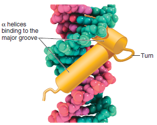
FIGURE 9.15 Key features of the structure of the double helix. Note: In the drawing on the left and in the inset, the planes of the bases and sugars are shown parallel to each other in order to depict the hydrogen bonding between the bases. In an actual DNA molecule, the bases would be rotated about 90° so the planes of the bases would be facing each other, as shown in Figure 9.16a.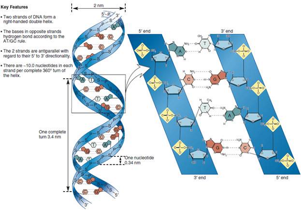
FIGURE 9.16 Two models of the double helix. (a) Ball-and-stick model of the double helix. The deoxyribose-phosphate backbone is shown in detail, whereas the bases are depicted as flattened rectangles.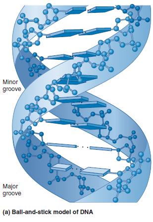
Explanation Verified
Like the helices present in DNA, there a...
Genetics: Analysis and Principles 4th Edition by Robert Brooker
Why don’t you like this exercise?
Other Minimum 8 character and maximum 255 character
Character 255