Deck 3: Protein Structure and Function
Full screen (f)
Question
Question
Question
Question
Question
Question
Question
Question
Question
Question
Question
Question
Question
Question
Question
Question
Question
Question
Question
Question
Question
Question
Question
Question
Question
Question
Question
Question
Question
Question
Question
Question
Question
Question
Question
Question
Question
Question
Question
Question
Question
Refer to the following paragraph and figure 3.1 to answer the following questions.
![<strong>Refer to the following paragraph and figure 3.1 to answer the following questions. Figure 3.1 Since structure correlates so well with function, biochemists are constantly looking for new ways to probe the complex structure of proteins in order to understand what they do and how they do it. One of the most powerful techniques in existence today is X-ray crystallography. The main difficulty with this technique is getting the protein to crystallize. Once crystallized, the protein is bombarded with X-rays to create a pattern that can be analyzed mathematically to determine the three-dimensional structure of the protein. This analysis has been performed by Krzysztof Palczewski on the protein rhodopsin, which is a light-sensitive protein found in species ranging from ancient bacteria (archaea)to humans. The structure (schematically shown above, where each letter represents an amino acid)is characterized by a single polypeptide chain with several α-helical segments that loop back and forth across the cell membrane. Another notable feature is the disulfide bond (-S-S-)that can be seen at the bottom of the third transmembrane segment. [Figure adapted from K. Palczewski et al., Science 289 (2000): 739.] How many times does the protein in Figure 3.1 cross the cell membrane?</strong> A)1 B)3 C)4 D)7 <div style=padding-top: 35px>](https://d2lvgg3v3hfg70.cloudfront.net/TB3733/11ea46a8_f568_afc5_95d4_292e393386ff_TB3733_00_TB3733_00_TB3733_00_TB3733_00_TB3733_00_TB3733_00.jpg)
Figure 3.1
Since structure correlates so well with function, biochemists are constantly looking for new ways to probe the complex structure of proteins in order to understand what they do and how they do it. One of the most powerful techniques in existence today is X-ray crystallography. The main difficulty with this technique is getting the protein to crystallize. Once crystallized, the protein is bombarded with X-rays to create a pattern that can be analyzed mathematically to determine the three-dimensional structure of the protein. This analysis has been performed by Krzysztof Palczewski on the protein rhodopsin, which is a light-sensitive protein found in species ranging from ancient bacteria (archaea)to humans. The structure (schematically shown above, where each letter represents an amino acid)is characterized by a single polypeptide chain with several α-helical segments that loop back and forth across the cell membrane. Another notable feature is the disulfide bond (-S-S-)that can be seen at the bottom of the third transmembrane segment. [Figure adapted from K. Palczewski et al., Science 289 (2000): 739.]
How many times does the protein in Figure 3.1 cross the cell membrane?
A)1
B)3
C)4
D)7
Question
Figure 3.2

Which of the following answers best describes the above metabolic pathway?
A)Anabolic;endergonic
B)Catabolic;endergonic
C)Anabolic;exergonic
D)Catabolic;exergonic
Which of the following answers best describes the above metabolic pathway?
A)Anabolic;endergonic
B)Catabolic;endergonic
C)Anabolic;exergonic
D)Catabolic;exergonic
Question
Question
Refer to the following paragraph and figure 3.1 to answer the following questions.
Figure 3.1
Since structure correlates so well with function, biochemists are constantly looking for new ways to probe the complex structure of proteins in order to understand what they do and how they do it. One of the most powerful techniques in existence today is X-ray crystallography. The main difficulty with this technique is getting the protein to crystallize. Once crystallized, the protein is bombarded with X-rays to create a pattern that can be analyzed mathematically to determine the three-dimensional structure of the protein. This analysis has been performed by Krzysztof Palczewski on the protein rhodopsin, which is a light-sensitive protein found in species ranging from ancient bacteria (archaea)to humans. The structure (schematically shown above, where each letter represents an amino acid)is characterized by a single polypeptide chain with several α-helical segments that loop back and forth across the cell membrane. Another notable feature is the disulfide bond (-S-S-)that can be seen at the bottom of the third transmembrane segment. [Figure adapted from K. Palczewski et al., Science 289 (2000): 739.]
If you were reading off the sequence of amino acids in Figure 3.1 to a biologist friend,what should the first three letters be?
A)M-N-G
B)A-P-A
C)It doesn't matter,since the protein has no polarity or directionality.
Question
Question
Question
Refer to the following paragraph and figure 3.1 to answer the following questions.
Figure 3.1
Since structure correlates so well with function, biochemists are constantly looking for new ways to probe the complex structure of proteins in order to understand what they do and how they do it. One of the most powerful techniques in existence today is X-ray crystallography. The main difficulty with this technique is getting the protein to crystallize. Once crystallized, the protein is bombarded with X-rays to create a pattern that can be analyzed mathematically to determine the three-dimensional structure of the protein. This analysis has been performed by Krzysztof Palczewski on the protein rhodopsin, which is a light-sensitive protein found in species ranging from ancient bacteria (archaea)to humans. The structure (schematically shown above, where each letter represents an amino acid)is characterized by a single polypeptide chain with several α-helical segments that loop back and forth across the cell membrane. Another notable feature is the disulfide bond (-S-S-)that can be seen at the bottom of the third transmembrane segment. [Figure adapted from K. Palczewski et al., Science 289 (2000): 739.]
Which term best describes the protein in Figure 3.1?
A)peripheral
B)external
C)internal
D)integral
Question
Which of the following metabolic pathways is Anabolic
A)
B)
C)
D)
A)
B)
C)
D)
Question
Refer to the following paragraph and figure 3.1 to answer the following questions.
Figure 3.1
Since structure correlates so well with function, biochemists are constantly looking for new ways to probe the complex structure of proteins in order to understand what they do and how they do it. One of the most powerful techniques in existence today is X-ray crystallography. The main difficulty with this technique is getting the protein to crystallize. Once crystallized, the protein is bombarded with X-rays to create a pattern that can be analyzed mathematically to determine the three-dimensional structure of the protein. This analysis has been performed by Krzysztof Palczewski on the protein rhodopsin, which is a light-sensitive protein found in species ranging from ancient bacteria (archaea)to humans. The structure (schematically shown above, where each letter represents an amino acid)is characterized by a single polypeptide chain with several α-helical segments that loop back and forth across the cell membrane. Another notable feature is the disulfide bond (-S-S-)that can be seen at the bottom of the third transmembrane segment. [Figure adapted from K. Palczewski et al., Science 289 (2000): 739.]
Refer to Figure 3.1.Which level of structure is being maintained by the disulfide bond?
A)primary
B)secondary
C)tertiary
D)quaternary
Question
Question
Refer to the following paragraph and figure 3.1 to answer the following questions.
Figure 3.1
Since structure correlates so well with function, biochemists are constantly looking for new ways to probe the complex structure of proteins in order to understand what they do and how they do it. One of the most powerful techniques in existence today is X-ray crystallography. The main difficulty with this technique is getting the protein to crystallize. Once crystallized, the protein is bombarded with X-rays to create a pattern that can be analyzed mathematically to determine the three-dimensional structure of the protein. This analysis has been performed by Krzysztof Palczewski on the protein rhodopsin, which is a light-sensitive protein found in species ranging from ancient bacteria (archaea)to humans. The structure (schematically shown above, where each letter represents an amino acid)is characterized by a single polypeptide chain with several α-helical segments that loop back and forth across the cell membrane. Another notable feature is the disulfide bond (-S-S-)that can be seen at the bottom of the third transmembrane segment. [Figure adapted from K. Palczewski et al., Science 289 (2000): 739.]
Identify the location of the disulfide bond in Figure 3.1.What is the name of the amino acids that are forming this bond?
A)cytosine
B)aspartic acid
C)cysteine
D)glycine
Question
Figure 3.4
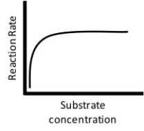
The figure above represents a reaction with a fixed enzyme concentration.As the concentration of substrate increases,the graph eventually levels off and reaches a plateau.Which of the following statements best describes the reason for this?
A)As the reaction proceeds,the enzyme is broken down by the substrate until no enzyme is left to continue the reaction.
B)As the reaction proceeds,all of the active sites of the enzyme become saturated such that a maximum catalytic rate is reached.
C)As the reaction proceeds,the product begins to occupy the active site of the enzyme such that a maximum catalytic rate is reached.
D)As the reaction proceeds,the enzyme is denatured due to the increased levels of product being formed.
Question
Refer to the following paragraph and figure 3.1 to answer the following questions.
Figure 3.1
Since structure correlates so well with function, biochemists are constantly looking for new ways to probe the complex structure of proteins in order to understand what they do and how they do it. One of the most powerful techniques in existence today is X-ray crystallography. The main difficulty with this technique is getting the protein to crystallize. Once crystallized, the protein is bombarded with X-rays to create a pattern that can be analyzed mathematically to determine the three-dimensional structure of the protein. This analysis has been performed by Krzysztof Palczewski on the protein rhodopsin, which is a light-sensitive protein found in species ranging from ancient bacteria (archaea)to humans. The structure (schematically shown above, where each letter represents an amino acid)is characterized by a single polypeptide chain with several α-helical segments that loop back and forth across the cell membrane. Another notable feature is the disulfide bond (-S-S-)that can be seen at the bottom of the third transmembrane segment. [Figure adapted from K. Palczewski et al., Science 289 (2000): 739.]
What is the location of the C-terminus of the protein in Figure 3.1?
A)extracellular
B)cytoplasm
C)embedded within the membrane
D)nucleus
Question
Figure 3.3
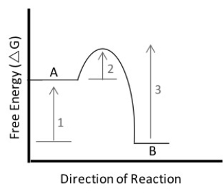
In the figure above,which of the following statements is true?
A)The reaction is considered to be endergonic.
B)The free energy change of the reaction is indicated by arrow #1.
C)The reaction will have a positive G value.
D)The free energy change of the reaction is indicated by arrow #2.
In the figure above,which of the following statements is true?
A)The reaction is considered to be endergonic.
B)The free energy change of the reaction is indicated by arrow #1.
C)The reaction will have a positive G value.
D)The free energy change of the reaction is indicated by arrow #2.

Unlock Deck
Sign up to unlock the cards in this deck!
Unlock Deck
Unlock Deck
1/54
Play
Full screen (f)
Deck 3: Protein Structure and Function
1
What prediction does the chemical evolution hypothesis make?
A)Nothing will happen,no new types of molecules will appear.
B)Molecules with carbon-carbon bonds will form.
C)Proteins will be produced before any other macromolecule.
D)A self-replicating macromolecule cannot exist because they all need interaction with another macromolecule.
A)Nothing will happen,no new types of molecules will appear.
B)Molecules with carbon-carbon bonds will form.
C)Proteins will be produced before any other macromolecule.
D)A self-replicating macromolecule cannot exist because they all need interaction with another macromolecule.
B
2
How does the structure of an amino acid enable it to play its most important roles in cells?
A)It can serve a wide variety of functions in a cell,because it contains the atoms most commonly found in organisms (C,H,N,and O).
B)Because both carboxyl and amino groups are present,polymerization is exergonic.In addition,the presence of a side chain makes the molecule water soluble.
C)The presence of carboxyl and amino groups gives it the ability to form peptide bonds,and its side chain gives it unique chemical properties.
D)Because each amino acid contains a variety of functional groups,they can participate in a wide variety of chemical reactions.
C
3
In solution,why do hydrolysis reactions occur more readily than condensation reactions?
A)Hydrolysis increases entropy and is exothermic.
B)Hydrolysis raises G,or Gibbs free energy.
C)Hydrolysis decreases entropy and is exothermic.
D)Hydrolysis increases entropy and is endothermic.
A)Hydrolysis increases entropy and is exothermic.
B)Hydrolysis raises G,or Gibbs free energy.
C)Hydrolysis decreases entropy and is exothermic.
D)Hydrolysis increases entropy and is endothermic.
A
4
Which of the following best describes the first living entity-the one responsible for the origin of life?
A)It was a monomer.
B)It was large and extremely complex.
C)It could make a copy of itself.
A)It was a monomer.
B)It was large and extremely complex.
C)It could make a copy of itself.
Unlock Deck
Unlock for access to all 54 flashcards in this deck.
Unlock Deck
k this deck
5
Which of the following involves an increase in entropy?
A)hydrolysis
B)condensation
C)polymerization
D)chemical evolution
A)hydrolysis
B)condensation
C)polymerization
D)chemical evolution
Unlock Deck
Unlock for access to all 54 flashcards in this deck.
Unlock Deck
k this deck
6
Which is not a role proteins play in organisms?
A)store genetic information
B)movement and shape changes
C)chemical signaling
D)structural support
A)store genetic information
B)movement and shape changes
C)chemical signaling
D)structural support
Unlock Deck
Unlock for access to all 54 flashcards in this deck.
Unlock Deck
k this deck
7
Three important functions of proteins are cell
A)wall composition,cushioning,and membrane fluidity.
B)movement,signaling,and reaction catalysis.
C)information coding,conversion,and transfer.
A)wall composition,cushioning,and membrane fluidity.
B)movement,signaling,and reaction catalysis.
C)information coding,conversion,and transfer.
Unlock Deck
Unlock for access to all 54 flashcards in this deck.
Unlock Deck
k this deck
8
In experiments that successfully simulate chemical evolution,why must at least some small,reduced molecules be present?
A)They act as proton donors in acid-base reactions.
B)They act as proton acceptors in acid-base reactions.
C)They act as electron acceptors in redox reactions.
D)They act as electron donors in redox reactions.
A)They act as proton donors in acid-base reactions.
B)They act as proton acceptors in acid-base reactions.
C)They act as electron acceptors in redox reactions.
D)They act as electron donors in redox reactions.
Unlock Deck
Unlock for access to all 54 flashcards in this deck.
Unlock Deck
k this deck
9
Suppose that Miller repeated his chemical evolution experiment,but without a source of electrical sparks.What would be the purpose?
A)to test if kinetic energy is required for chemical evolution
B)to test the hypothesis that reduced molecules are required for chemical evolution
C)to test the hypothesis that both reduced molecules and kinetic energy are required for chemical evolution
D)to make sure that the glassware had not been contaminated and that any new molecules found were actually produced by chemical evolution
Unlock Deck
Unlock for access to all 54 flashcards in this deck.
Unlock Deck
k this deck
10
Which one of the following is not a component of each monomer used to make proteins?
A)a phosphorous atom,P
B)an amino functional group,NH₂
C)a side chain,R
D)a carboxyl group,COOH
A)a phosphorous atom,P
B)an amino functional group,NH₂
C)a side chain,R
D)a carboxyl group,COOH
Unlock Deck
Unlock for access to all 54 flashcards in this deck.
Unlock Deck
k this deck
11
In interstellar space,millions of ice-encrusted dust particles contain simple carbon-containing compounds.When particles like these are exposed to solar radiation,more complex organic molecules form on the surfaces of the dust.What is the significance of these findings?
A)Chemical evolution occurs only in outer space and was not possible on Earth.
B)Life began in outer space.
C)Life exists in outer space.
D)Chemical evolution occurs readily in outer space.
A)Chemical evolution occurs only in outer space and was not possible on Earth.
B)Life began in outer space.
C)Life exists in outer space.
D)Chemical evolution occurs readily in outer space.
Unlock Deck
Unlock for access to all 54 flashcards in this deck.
Unlock Deck
k this deck
12
At the pH found in cells (about 7.0),what happens to the amino group on an amino acid?
A)It acts as a base and gains a proton,giving it a positive charge.
B)It acts as an acid and loses a proton,giving it a negative charge.
C)It is reduced and tends to act as an electron donor in redox reactions.
D)It remains neutral,like water,and does not have a charge.
A)It acts as a base and gains a proton,giving it a positive charge.
B)It acts as an acid and loses a proton,giving it a negative charge.
C)It is reduced and tends to act as an electron donor in redox reactions.
D)It remains neutral,like water,and does not have a charge.
Unlock Deck
Unlock for access to all 54 flashcards in this deck.
Unlock Deck
k this deck
13
Suppose you discovered a new amino acid.Its R-group contains only hydrogen and carbon atoms.Predict the behavior of this amino acid.
A)It is hydrophobic.
B)It is hydrophilic.
C)Relative to the amino acids found in organisms,its interactions with water will be intermediate.
D)Relative to the amino acids found in organisms,its interactions with water will be very high.
A)It is hydrophobic.
B)It is hydrophilic.
C)Relative to the amino acids found in organisms,its interactions with water will be intermediate.
D)Relative to the amino acids found in organisms,its interactions with water will be very high.
Unlock Deck
Unlock for access to all 54 flashcards in this deck.
Unlock Deck
k this deck
14
An isomer of a particular molecule is
A)a molecule that has the same structure as the target molecule,but a different formula.
B)a molecule that is the same except it has an additional side group.
C)another copy of the same molecule.
D)a molecule that has the same formula,but a different structure.
A)a molecule that has the same structure as the target molecule,but a different formula.
B)a molecule that is the same except it has an additional side group.
C)another copy of the same molecule.
D)a molecule that has the same formula,but a different structure.
Unlock Deck
Unlock for access to all 54 flashcards in this deck.
Unlock Deck
k this deck
15
Consider the experiment that Stanley Miller did to simulate chemical evolution.Recall that a glass flask held the reduced gases NH₃,CH₄,and H₂ and that the gases were exposed to electrical sparks.What is the null hypothesis in the experiment?
A)Chemical evolution does not occur.
B)Chemical evolution requires the presence of reduced molecules.
C)Chemical evolution requires continuous heating.
D)Chemical evolution requires a source of kinetic energy.
E)Chemical evolution occurs only on Earth.
A)Chemical evolution does not occur.
B)Chemical evolution requires the presence of reduced molecules.
C)Chemical evolution requires continuous heating.
D)Chemical evolution requires a source of kinetic energy.
E)Chemical evolution occurs only on Earth.
Unlock Deck
Unlock for access to all 54 flashcards in this deck.
Unlock Deck
k this deck
16
At the pH found in cells (about 7.0),what happens to the carboxyl group on an amino acid?
A)It acts as a base and gains a proton,giving it a positive charge.
B)It acts as an acid and loses a proton,giving it a negative charge.
C)It is oxidized and tends to act as an electron acceptor in redox reactions.
D)It remains neutral,like water,and does not have a charge.
A)It acts as a base and gains a proton,giving it a positive charge.
B)It acts as an acid and loses a proton,giving it a negative charge.
C)It is oxidized and tends to act as an electron acceptor in redox reactions.
D)It remains neutral,like water,and does not have a charge.
Unlock Deck
Unlock for access to all 54 flashcards in this deck.
Unlock Deck
k this deck
17
Why are polymerization reactions endergonic?
A)They reduce entropy.
B)They release heat,making the reactant monomers move faster.
C)Because the condensation and hydrolysis reactions are equally spontaneous.
D)Because polymers are energetically more stable and have lower potential energy than monomers do.
A)They reduce entropy.
B)They release heat,making the reactant monomers move faster.
C)Because the condensation and hydrolysis reactions are equally spontaneous.
D)Because polymers are energetically more stable and have lower potential energy than monomers do.
Unlock Deck
Unlock for access to all 54 flashcards in this deck.
Unlock Deck
k this deck
18
What is the pattern component of the theory of chemical evolution?
A)Both heat and electrical discharges are required for chemical evolution to occur.
B)Most chemical evolution occurred at black smokers.
C)The process occurred at black smokers,in the atmosphere and oceans,and in outer space.
D)Increasingly complex carbon-containing molecules formed early in Earth history.
Unlock Deck
Unlock for access to all 54 flashcards in this deck.
Unlock Deck
k this deck
19
Amino acid side chains (R groups)with what characteristic(s)dissolve best in water?
A)small sizes and simple structures
B)at least one ring structure
C)polarity or charged structures
D)the presence of sulfur
A)small sizes and simple structures
B)at least one ring structure
C)polarity or charged structures
D)the presence of sulfur
Unlock Deck
Unlock for access to all 54 flashcards in this deck.
Unlock Deck
k this deck
20
What is the process component of the theory of chemical evolution?
A)Acid-base reactions resulted in the formation of large,complex organic molecules.
B)Kinetic energy was transformed into chemical energy.
C)During polymerization reactions,hydrolysis competed with condensation.
D)The process occurred at black smokers,in the atmosphere and oceans,and in outer space.
Unlock Deck
Unlock for access to all 54 flashcards in this deck.
Unlock Deck
k this deck
21
HIV is the virus that causes AIDS.In the mid-1990s,researchers discovered an enzyme in HIV called protease.Once the enzyme's structure was known,researchers began looking for drugs that would fit into the active site and block it.If this strategy for stopping HIV infections were successful,it would be an example of what phenomenon?
A)vaccination
B)poisoning
C)allosteric regulation
D)competitive inhibition
A)vaccination
B)poisoning
C)allosteric regulation
D)competitive inhibition
Unlock Deck
Unlock for access to all 54 flashcards in this deck.
Unlock Deck
k this deck
22
Consider the HIV enzyme called protease.The amino acid residues at the active site are highly hydrophobic.In designing a drug that would bind to the active site and jam it,researchers should use which type of molecule?
A)hydrophobic
B)polar
C)charged
D)acidic
A)hydrophobic
B)polar
C)charged
D)acidic
Unlock Deck
Unlock for access to all 54 flashcards in this deck.
Unlock Deck
k this deck
23
An enzyme has a total of four active sites.When you denature the molecule and study its composition,you find that each active site occurs on a different polypeptide.Which of the following hypotheses does this observation support?
A)The enzyme is subject to allosteric regulation.
B)The enzyme requires a cofactor to function normally.
C)The protein's structure is affected by temperature and pH.
D)The protein has quaternary structure.
A)The enzyme is subject to allosteric regulation.
B)The enzyme requires a cofactor to function normally.
C)The protein's structure is affected by temperature and pH.
D)The protein has quaternary structure.
Unlock Deck
Unlock for access to all 54 flashcards in this deck.
Unlock Deck
k this deck
24
If the primary structure of a protein is incorrect
A)the secondary structure will be correct.
B)the tertiary structure will be correct.
C)any quaternary structure will be correct.
D)any higher-level folding of the protein will be incorrect.
A)the secondary structure will be correct.
B)the tertiary structure will be correct.
C)any quaternary structure will be correct.
D)any higher-level folding of the protein will be incorrect.
Unlock Deck
Unlock for access to all 54 flashcards in this deck.
Unlock Deck
k this deck
25
Which of the following observations is the strongest argument in favor of the hypothesis that protein structure and function are correlated?
A)Proteins function best at certain temperatures.
B)Proteins have four distinct levels of structure and many functions.
C)Enzymes tend to be globular in shape.
D)Denatured (unfolded)proteins do not function normally.
A)Proteins function best at certain temperatures.
B)Proteins have four distinct levels of structure and many functions.
C)Enzymes tend to be globular in shape.
D)Denatured (unfolded)proteins do not function normally.
Unlock Deck
Unlock for access to all 54 flashcards in this deck.
Unlock Deck
k this deck
26
When polymerization of a protein is complete,but the protein is still completely linear,what is the highest level of structure that has been completed?
A)primary
B)secondary
C)tertiary
D)quaternary
A)primary
B)secondary
C)tertiary
D)quaternary
Unlock Deck
Unlock for access to all 54 flashcards in this deck.
Unlock Deck
k this deck
27
In cells,the activity of enzymes is often regulated by other molecules.Why is this necessary?
A)because all enzymes require some help from another molecule to function correctly
B)because other molecules are necessary to prevent enzymes from denaturing
C)because each enzyme has multiple functions
D)because it is unlikely that all reaction products are required all of the time
A)because all enzymes require some help from another molecule to function correctly
B)because other molecules are necessary to prevent enzymes from denaturing
C)because each enzyme has multiple functions
D)because it is unlikely that all reaction products are required all of the time
Unlock Deck
Unlock for access to all 54 flashcards in this deck.
Unlock Deck
k this deck
28
A peptide bond is
A)an ionic bond,not a covalent one.
B)a triple covalent bond.
C)a particularly stable,planar covalent bond.
D)a particularly unstable covalent bond.
A)an ionic bond,not a covalent one.
B)a triple covalent bond.
C)a particularly stable,planar covalent bond.
D)a particularly unstable covalent bond.
Unlock Deck
Unlock for access to all 54 flashcards in this deck.
Unlock Deck
k this deck
29
Which of the following would be an example of a cofactor?
A)an enzyme active site that contains an α-helix
B)the nonprotein heme group in a hemoglobin molecule
C)the disulfide bridge that forms between cysteine residues
D)a β-pleated sheet hidden on the inside of a protein's tertiary structure
A)an enzyme active site that contains an α-helix
B)the nonprotein heme group in a hemoglobin molecule
C)the disulfide bridge that forms between cysteine residues
D)a β-pleated sheet hidden on the inside of a protein's tertiary structure
Unlock Deck
Unlock for access to all 54 flashcards in this deck.
Unlock Deck
k this deck
30
You are studying a protein that is shaped like a doughnut.The shape is a function of which level(s)of protein structure?
A)primary only
B)secondary only
C)tertiary only
D)secondary and tertiary only
E)primary,secondary,and tertiary
A)primary only
B)secondary only
C)tertiary only
D)secondary and tertiary only
E)primary,secondary,and tertiary
Unlock Deck
Unlock for access to all 54 flashcards in this deck.
Unlock Deck
k this deck
31
Recent technological advances have made it more feasible than ever to work out the three-dimensional structure of proteins.There is intense interest in this research field,called structural biology.Why?
A)Understanding structure should help us understand function.
B)Understanding structure can help in the design of drugs that alter the function of certain proteins.
C)Solving a protein's 3-D structure can lead to a better understanding of how the molecule works-for example,by identifying the active site and determining if there are regulatory sites.
D)All of the above apply.
Unlock Deck
Unlock for access to all 54 flashcards in this deck.
Unlock Deck
k this deck
32
What type of interaction is directly responsible for the formation of secondary structure?
A)peptide bonds between adjacent amino acids
B)peptide bonds between nonadjacent amino acids
C)hydrogen bonds between sections of the polypeptide backbone
D)hydrogen bonds between side chains of amino acids
A)peptide bonds between adjacent amino acids
B)peptide bonds between nonadjacent amino acids
C)hydrogen bonds between sections of the polypeptide backbone
D)hydrogen bonds between side chains of amino acids
Unlock Deck
Unlock for access to all 54 flashcards in this deck.
Unlock Deck
k this deck
33
When,and by whom,was the lock-and-key model of enzyme specificity developed?
A)1894 by Fischer
B)1894 by Miller
C)1936 by Fischer
D)1956 by Fischer
E)1974 by Haldane and Oparin
A)1894 by Fischer
B)1894 by Miller
C)1936 by Fischer
D)1956 by Fischer
E)1974 by Haldane and Oparin
Unlock Deck
Unlock for access to all 54 flashcards in this deck.
Unlock Deck
k this deck
34
Which of the following is not true when comparing an uncatalyzed reaction to the same reaction with a catalyst?
A)The catalyzed reaction will be faster.
B)The catalyzed reaction will have a different ∆G.
C)The catalyzed reaction will have lower activation energy.
D)The catalyzed reaction will not consume any of the catalyst.
A)The catalyzed reaction will be faster.
B)The catalyzed reaction will have a different ∆G.
C)The catalyzed reaction will have lower activation energy.
D)The catalyzed reaction will not consume any of the catalyst.
Unlock Deck
Unlock for access to all 54 flashcards in this deck.
Unlock Deck
k this deck
35
You've just sequenced a new protein found in mice and observe that sulfur-containing cysteine residues occur at regular intervals.What is the significance of this finding?
A)Cysteine residues are required for the formation of α-helices and β-pleated sheets.
B)It will be important to include cysteine in the diet of the mice.
C)Cysteine residues are involved in disulfide bridges that help form tertiary structure.
D)Cysteine causes bends,or angles,to occur in the tertiary structure of proteins.
Unlock Deck
Unlock for access to all 54 flashcards in this deck.
Unlock Deck
k this deck
36
You have isolated a previously unstudied protein,identified its complete structure in detail,and determined that it catalyzes the breakdown of a large substrate.You notice it has two binding sites.One of these is large,apparently the bonding site for the large substrate;the other is small,possibly a binding site for a regulatory molecule.What do these findings tell you about the mechanism of this protein?
A)It is probably a structural protein that is involved in cell-to-cell adhesion.
B)It is probably an enzyme that works through allosteric regulation.
C)It is probably an enzyme that works through competitive inhibition.
D)It is probably a cell membrane transport protein,like an ion channel.
E)It is probably a structural protein found in cartilage or skeletal tissue.
Unlock Deck
Unlock for access to all 54 flashcards in this deck.
Unlock Deck
k this deck
37
You've discovered an enzyme that can catalyze two different chemical reactions.Which of the following is most likely to be correct?
A)The enzyme contains both α-helices and β-pleated sheets.
B)The enzyme is subject to both competitive inhibition and allosteric regulation.
C)Two types of allosteric regulation occur-the binding of one molecule activates the enzyme,while the binding of a different molecule inhibits it.
D)Either the enzyme has two distinct active sites,or the reactants involved in the two reactions are very similar in size and shape.
Unlock Deck
Unlock for access to all 54 flashcards in this deck.
Unlock Deck
k this deck
38
A series of hydrophobic side chains will congregate together as a protein folds in an aqueous solution and be stabilized by
A)disulfide bonds.
B)van der Waals interaction.
C)hydrogen bonds.
D)quaternary structure bonds.
A)disulfide bonds.
B)van der Waals interaction.
C)hydrogen bonds.
D)quaternary structure bonds.
Unlock Deck
Unlock for access to all 54 flashcards in this deck.
Unlock Deck
k this deck
39
Which of the following best describes primary structure in proteins?
A)It is the number of amino acids present in the complete protein.
B)It is the number of peptide bonds in the complete protein.
C)It is the sequence of amino acids in the complete protein.
D)It is the number of α-helices and β-pleated sheets in the complete protein.
A)It is the number of amino acids present in the complete protein.
B)It is the number of peptide bonds in the complete protein.
C)It is the sequence of amino acids in the complete protein.
D)It is the number of α-helices and β-pleated sheets in the complete protein.
Unlock Deck
Unlock for access to all 54 flashcards in this deck.
Unlock Deck
k this deck
40
Several of the molecules called vitamins act as enzyme cofactors.Vitamin deficiencies cause disease.What is the most direct explanation for this?
A)Vitamins combine with nonprotein molecules to delay the onset of disease.
B)If cofactors are missing,enzymes cannot function properly,and important reaction products will be absent from cells.
C)Normal regulation cannot occur in the absence of cofactors.As a result,all enzymes will function all of the time.
D)Cofactors inhibit enzymes found in disease-causing bacteria and viruses.When cofactors are absent,these disease-causing agents multiply.
Unlock Deck
Unlock for access to all 54 flashcards in this deck.
Unlock Deck
k this deck
41
Refer to the following paragraph and figure 3.1 to answer the following questions.
Figure 3.1
Since structure correlates so well with function, biochemists are constantly looking for new ways to probe the complex structure of proteins in order to understand what they do and how they do it. One of the most powerful techniques in existence today is X-ray crystallography. The main difficulty with this technique is getting the protein to crystallize. Once crystallized, the protein is bombarded with X-rays to create a pattern that can be analyzed mathematically to determine the three-dimensional structure of the protein. This analysis has been performed by Krzysztof Palczewski on the protein rhodopsin, which is a light-sensitive protein found in species ranging from ancient bacteria (archaea)to humans. The structure (schematically shown above, where each letter represents an amino acid)is characterized by a single polypeptide chain with several α-helical segments that loop back and forth across the cell membrane. Another notable feature is the disulfide bond (-S-S-)that can be seen at the bottom of the third transmembrane segment. [Figure adapted from K. Palczewski et al., Science 289 (2000): 739.]
How many times does the protein in Figure 3.1 cross the cell membrane?
A)1
B)3
C)4
D)7
Unlock Deck
Unlock for access to all 54 flashcards in this deck.
Unlock Deck
k this deck
42
Figure 3.2
Which of the following answers best describes the above metabolic pathway?
A)Anabolic;endergonic
B)Catabolic;endergonic
C)Anabolic;exergonic
D)Catabolic;exergonic
Which of the following answers best describes the above metabolic pathway?
A)Anabolic;endergonic
B)Catabolic;endergonic
C)Anabolic;exergonic
D)Catabolic;exergonic
Unlock Deck
Unlock for access to all 54 flashcards in this deck.
Unlock Deck
k this deck
43
Which one of the following is not a component of each monomer used to make proteins?
A)an iron atom,Fe
B)an amino functional group,NH₂
C)a side chain,R
D)a carboxyl group,COOH
A)an iron atom,Fe
B)an amino functional group,NH₂
C)a side chain,R
D)a carboxyl group,COOH
Unlock Deck
Unlock for access to all 54 flashcards in this deck.
Unlock Deck
k this deck
44
Refer to the following paragraph and figure 3.1 to answer the following questions.
Figure 3.1
Since structure correlates so well with function, biochemists are constantly looking for new ways to probe the complex structure of proteins in order to understand what they do and how they do it. One of the most powerful techniques in existence today is X-ray crystallography. The main difficulty with this technique is getting the protein to crystallize. Once crystallized, the protein is bombarded with X-rays to create a pattern that can be analyzed mathematically to determine the three-dimensional structure of the protein. This analysis has been performed by Krzysztof Palczewski on the protein rhodopsin, which is a light-sensitive protein found in species ranging from ancient bacteria (archaea)to humans. The structure (schematically shown above, where each letter represents an amino acid)is characterized by a single polypeptide chain with several α-helical segments that loop back and forth across the cell membrane. Another notable feature is the disulfide bond (-S-S-)that can be seen at the bottom of the third transmembrane segment. [Figure adapted from K. Palczewski et al., Science 289 (2000): 739.]
If you were reading off the sequence of amino acids in Figure 3.1 to a biologist friend,what should the first three letters be?
A)M-N-G
B)A-P-A
C)It doesn't matter,since the protein has no polarity or directionality.
Unlock Deck
Unlock for access to all 54 flashcards in this deck.
Unlock Deck
k this deck
45
You are studying reaction X.Which of the following scenarios would likely NOT help you to increase the rate of this reaction?
A)Adding more product to an excess of substrate.
B)Adding more enzyme to an excess of substrate.
C)Adding more substrate to an excess of enzyme.
A)Adding more product to an excess of substrate.
B)Adding more enzyme to an excess of substrate.
C)Adding more substrate to an excess of enzyme.
Unlock Deck
Unlock for access to all 54 flashcards in this deck.
Unlock Deck
k this deck
46
The aquaporin family of proteins plays a major role in the transport of water all over the body.During the folding process of these proteins,α-helices start forming as
A)part of the primary structure of the protein.
B)part of the secondary structure of the protein.
C)part of the tertiary structure of the protein.
D)part of the quaternary structure of the protein.
A)part of the primary structure of the protein.
B)part of the secondary structure of the protein.
C)part of the tertiary structure of the protein.
D)part of the quaternary structure of the protein.
Unlock Deck
Unlock for access to all 54 flashcards in this deck.
Unlock Deck
k this deck
47
Refer to the following paragraph and figure 3.1 to answer the following questions.
Figure 3.1
Since structure correlates so well with function, biochemists are constantly looking for new ways to probe the complex structure of proteins in order to understand what they do and how they do it. One of the most powerful techniques in existence today is X-ray crystallography. The main difficulty with this technique is getting the protein to crystallize. Once crystallized, the protein is bombarded with X-rays to create a pattern that can be analyzed mathematically to determine the three-dimensional structure of the protein. This analysis has been performed by Krzysztof Palczewski on the protein rhodopsin, which is a light-sensitive protein found in species ranging from ancient bacteria (archaea)to humans. The structure (schematically shown above, where each letter represents an amino acid)is characterized by a single polypeptide chain with several α-helical segments that loop back and forth across the cell membrane. Another notable feature is the disulfide bond (-S-S-)that can be seen at the bottom of the third transmembrane segment. [Figure adapted from K. Palczewski et al., Science 289 (2000): 739.]
Which term best describes the protein in Figure 3.1?
A)peripheral
B)external
C)internal
D)integral
Unlock Deck
Unlock for access to all 54 flashcards in this deck.
Unlock Deck
k this deck
48
Which of the following metabolic pathways is Anabolic
A)
B)
C)
D)
A)
B)
C)
D)
Unlock Deck
Unlock for access to all 54 flashcards in this deck.
Unlock Deck
k this deck
49
Refer to the following paragraph and figure 3.1 to answer the following questions.
Figure 3.1
Since structure correlates so well with function, biochemists are constantly looking for new ways to probe the complex structure of proteins in order to understand what they do and how they do it. One of the most powerful techniques in existence today is X-ray crystallography. The main difficulty with this technique is getting the protein to crystallize. Once crystallized, the protein is bombarded with X-rays to create a pattern that can be analyzed mathematically to determine the three-dimensional structure of the protein. This analysis has been performed by Krzysztof Palczewski on the protein rhodopsin, which is a light-sensitive protein found in species ranging from ancient bacteria (archaea)to humans. The structure (schematically shown above, where each letter represents an amino acid)is characterized by a single polypeptide chain with several α-helical segments that loop back and forth across the cell membrane. Another notable feature is the disulfide bond (-S-S-)that can be seen at the bottom of the third transmembrane segment. [Figure adapted from K. Palczewski et al., Science 289 (2000): 739.]
Refer to Figure 3.1.Which level of structure is being maintained by the disulfide bond?
A)primary
B)secondary
C)tertiary
D)quaternary
Unlock Deck
Unlock for access to all 54 flashcards in this deck.
Unlock Deck
k this deck
50
The aquaporin family of proteins plays a major role in the transport of water all over the body.During the folding process of these proteins,α-helices start forming as
A)part of the primary structure of the protein.
B)part of the secondary structure of the protein.
C)part of the tertiary structure of the protein.
D)part of the quaternary structure of the protein.
A)part of the primary structure of the protein.
B)part of the secondary structure of the protein.
C)part of the tertiary structure of the protein.
D)part of the quaternary structure of the protein.
Unlock Deck
Unlock for access to all 54 flashcards in this deck.
Unlock Deck
k this deck
51
Refer to the following paragraph and figure 3.1 to answer the following questions.
Figure 3.1
Since structure correlates so well with function, biochemists are constantly looking for new ways to probe the complex structure of proteins in order to understand what they do and how they do it. One of the most powerful techniques in existence today is X-ray crystallography. The main difficulty with this technique is getting the protein to crystallize. Once crystallized, the protein is bombarded with X-rays to create a pattern that can be analyzed mathematically to determine the three-dimensional structure of the protein. This analysis has been performed by Krzysztof Palczewski on the protein rhodopsin, which is a light-sensitive protein found in species ranging from ancient bacteria (archaea)to humans. The structure (schematically shown above, where each letter represents an amino acid)is characterized by a single polypeptide chain with several α-helical segments that loop back and forth across the cell membrane. Another notable feature is the disulfide bond (-S-S-)that can be seen at the bottom of the third transmembrane segment. [Figure adapted from K. Palczewski et al., Science 289 (2000): 739.]
Identify the location of the disulfide bond in Figure 3.1.What is the name of the amino acids that are forming this bond?
A)cytosine
B)aspartic acid
C)cysteine
D)glycine
Unlock Deck
Unlock for access to all 54 flashcards in this deck.
Unlock Deck
k this deck
52
Figure 3.4
The figure above represents a reaction with a fixed enzyme concentration.As the concentration of substrate increases,the graph eventually levels off and reaches a plateau.Which of the following statements best describes the reason for this?
A)As the reaction proceeds,the enzyme is broken down by the substrate until no enzyme is left to continue the reaction.
B)As the reaction proceeds,all of the active sites of the enzyme become saturated such that a maximum catalytic rate is reached.
C)As the reaction proceeds,the product begins to occupy the active site of the enzyme such that a maximum catalytic rate is reached.
D)As the reaction proceeds,the enzyme is denatured due to the increased levels of product being formed.
Unlock Deck
Unlock for access to all 54 flashcards in this deck.
Unlock Deck
k this deck
53
Refer to the following paragraph and figure 3.1 to answer the following questions.
Figure 3.1
Since structure correlates so well with function, biochemists are constantly looking for new ways to probe the complex structure of proteins in order to understand what they do and how they do it. One of the most powerful techniques in existence today is X-ray crystallography. The main difficulty with this technique is getting the protein to crystallize. Once crystallized, the protein is bombarded with X-rays to create a pattern that can be analyzed mathematically to determine the three-dimensional structure of the protein. This analysis has been performed by Krzysztof Palczewski on the protein rhodopsin, which is a light-sensitive protein found in species ranging from ancient bacteria (archaea)to humans. The structure (schematically shown above, where each letter represents an amino acid)is characterized by a single polypeptide chain with several α-helical segments that loop back and forth across the cell membrane. Another notable feature is the disulfide bond (-S-S-)that can be seen at the bottom of the third transmembrane segment. [Figure adapted from K. Palczewski et al., Science 289 (2000): 739.]
What is the location of the C-terminus of the protein in Figure 3.1?
A)extracellular
B)cytoplasm
C)embedded within the membrane
D)nucleus
Unlock Deck
Unlock for access to all 54 flashcards in this deck.
Unlock Deck
k this deck
54
Figure 3.3
In the figure above,which of the following statements is true?
A)The reaction is considered to be endergonic.
B)The free energy change of the reaction is indicated by arrow #1.
C)The reaction will have a positive G value.
D)The free energy change of the reaction is indicated by arrow #2.
In the figure above,which of the following statements is true?
A)The reaction is considered to be endergonic.
B)The free energy change of the reaction is indicated by arrow #1.
C)The reaction will have a positive G value.
D)The free energy change of the reaction is indicated by arrow #2.
Unlock Deck
Unlock for access to all 54 flashcards in this deck.
Unlock Deck
k this deck
Unlock Deck
Unlock for access to all 54 flashcards in this deck.